Table of Contents >> Show >> Hide
- What Is an Ultrasound?
- Why Your Doctor Might Order an Ultrasound
- Common Types of Ultrasound Exams
- Is Ultrasound Safe?
- What Happens During an Ultrasound Procedure?
- How to Prepare for an Ultrasound
- What Ultrasound Results Can and Can’t Tell You
- Real-Life Ultrasound Experiences: What It Feels Like and Practical Tips
- When to Call Your Doctor After an Ultrasound
- The Bottom Line
If you’ve been told you need an ultrasound, you might picture a cool black-and-white screen, some mysterious whooshing sounds, and a generous amount of cold, squishy gel. Good news: you’re not far off. Ultrasound is one of the most common and safest imaging tests doctors use to peek inside your body without a single incision or dose of radiation. Think of it as a high-tech echo that turns sound into pictures.
In this guide, we’ll walk through what an ultrasound actually is, why doctors order it, what happens during the procedure, how to prepare (yes, sometimes that means fasting or holding a very full bladder), and what to expect afterward. By the end, you’ll know exactly what’s going on and can show up for your scan feeling informed, prepared, and a whole lot calmer.
What Is an Ultrasound?
At its core, an ultrasound (also called sonography or ultrasonography) is an imaging test that uses high-frequency sound waves to create real-time pictures or video of the inside of your body. A small handheld device called a transducer sends sound waves into your body and listens for the echoes as they bounce off organs, blood vessels, or a developing baby. A computer turns those echoes into an image called a sonogram.
Unlike X-rays or CT scans, ultrasound uses non-ionizing sound energy, not radiation. That’s a big reason it’s a go-to test during pregnancy and for repeated imaging over time.
How ultrasound works (in plain English)
You can think of ultrasound like yelling into a canyon and listening for the echoexcept much more polite and highly trained. The transducer:
- Sends out sound waves at a frequency too high for humans to hear.
- Waits a split second for the waves to bounce back from tissues of different densities.
- Sends those returning echoes to a computer, which converts them into moving images.
Different tissues (like fluid, soft tissue, and bone) reflect sound differently. That’s why organs, blood flow, and babies show up with different shades and patterns on the screen.
Why Your Doctor Might Order an Ultrasound
Ultrasound is surprisingly versatile. Doctors use it for both diagnosis and monitoring. Common reasons include:
- Investigating symptoms such as abdominal pain, pelvic pain, swelling, or a lump.
- Evaluating organs like the liver, gallbladder, kidneys, pancreas, bladder, uterus, ovaries, and thyroid.
- Monitoring pregnancy to check the baby’s growth, heartbeat, position, and certain structures like the placenta.
- Checking blood flow with Doppler ultrasound to look for clots, narrowed arteries, or vein problems.
- Guiding procedures such as needle biopsies, fluid drainage, or injections into joints or cysts.
- Assessing the heart (echocardiogram) to look at valves, pumping function, and overall structure.
In many cases, ultrasound is the first imaging test your provider orders because it’s safe, quick, and relatively affordable compared with other scans.
Common Types of Ultrasound Exams
Abdominal ultrasound
This scan focuses on organs in your abdomensuch as the liver, gallbladder, pancreas, spleen, kidneys, and sometimes major blood vessels. It’s often used to look for gallstones, liver disease, kidney stones, or causes of upper belly pain and bloating.
Pelvic and obstetric ultrasound
A pelvic ultrasound examines the uterus, ovaries, fallopian tubes, bladder, and nearby structures. In people who are pregnant, it becomes an obstetric ultrasound and is used to check on the fetus, confirm due dates, and look for certain developmental or structural concerns.
Pelvic ultrasounds can be done:
- Transabdominally (over the lower abdomen with a full bladder).
- Transvaginally (using a thin, covered probe placed gently into the vagina for a closer view of pelvic organs).
Vascular (Doppler) ultrasound
Vascular ultrasound uses special techniques (Doppler) to show blood moving through arteries and veins. It’s commonly used to look for blood clots, narrowed arteries in the neck or legs, or vein valve problems that cause leg swelling and varicose veins.
Echocardiogram (heart ultrasound)
An echocardiogram is an ultrasound of your heart. It shows how well the heart muscle pumps, whether the valves open and close properly, and if there are any structural problems. It’s a key test for heart failure, valve disease, and many other cardiac conditions.
Other specialized ultrasounds
Depending on your symptoms, your provider might order:
- Thyroid ultrasound to evaluate nodules or enlargement in the neck.
- Breast ultrasound to look at a lump or clarify findings from a mammogram.
- Musculoskeletal ultrasound to examine tendons, ligaments, and joints.
- Kidney or bladder ultrasound for urinary symptoms or blood in the urine.
Is Ultrasound Safe?
This is often the first question people askand it’s a reasonable one. Ultrasound has been used in medicine for decades and has an excellent safety record when performed properly by trained professionals. Because it doesn’t use ionizing radiation, it doesn’t carry the same long-term radiation risks associated with X-rays or CT scans.
That said, “safe” in medicine usually comes with an asterisk: use it when it’s medically needed. Professional organizations and regulators emphasize that ultrasounds should be ordered for clear clinical reasons, not just for nonmedical keepsake videos or frequent scans “just for fun.”
Possible downsides are usually minor and can include:
- Temporary discomfort if the technologist has to press firmly on a sore area.
- Some awkwardness during internal exams such as transvaginal or transrectal ultrasound.
- False alarms or unclear findings that lead to more tests or follow-up imaging.
Overall, though, ultrasound is considered one of the safest imaging methods we haveespecially valuable in pregnancy and in children, where avoiding radiation is a big priority.
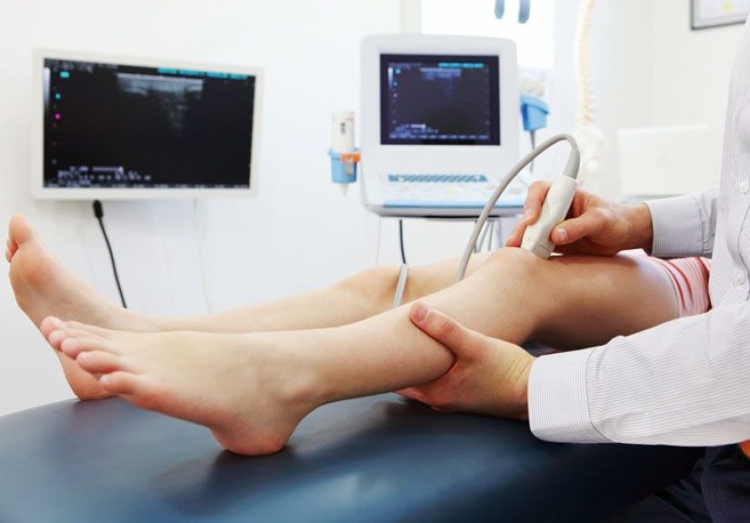
What Happens During an Ultrasound Procedure?
While the exact details depend on the type of ultrasound, most external exams follow a similar script. Here’s what typically happens:
- Check in and review your history. You’ll arrive, check in, and may be asked about your symptoms, medications, allergies, and prior imaging tests.
- Change clothes if needed. For many scans, you’ll wear a gown or loosen clothing around the area being examined. You may be asked to remove jewelry or piercings near the site.
- Get positioned on the exam table. You’ll lie on your back, side, or occasionally sit up, depending on what’s being imaged.
-
Gel time. The technologist (often a diagnostic medical sonographer) spreads a water-based gel on your skin. This gel feels cool and a bit sticky, but it:
- Helps the transducer glide smoothly over your skin.
- Pushes out tiny air pockets that would block sound waves.
- Scanning. The technologist moves the transducer over the area, sometimes pressing more firmly or asking you to hold your breath, roll to one side, or change positions. You might see the images on a screen in real time.
- Internal exams (if needed). For certain scans (like transvaginal or transrectal ultrasounds), a specially shaped probe covered with a protective sheath and gel is gently inserted. These exams are usually short and should not be painful, though they can feel uncomfortable or awkward.
- Wrap-up. Once the technologist has the images they need, they’ll wipe off the gel, help you sit up, and you can get dressed. A radiologist or other physician will later review the images and send a report to your ordering provider.
How to Prepare for an Ultrasound
Preparation depends on the type of ultrasound you’re having. Your doctor or imaging center should give you specific instructionsthose always take priority. But here are common scenarios so you know what to expect.
Preparing for an abdominal ultrasound
For many abdominal scans (such as looking at the liver, gallbladder, or pancreas), you’ll be asked to:
- Fast for 8–12 hours before the exam (no food and sometimes no drinks except small sips of water).
- Avoid certain foods that cause gas the day before, if instructed (like carbonated drinks or very fatty meals).
Why the hunger games? Food and fluid in your stomach and intestines can create gas and obscure the view, especially of the gallbladder and nearby structures. Fasting helps produce clearer, more accurate images.
Preparing for pelvic or obstetric ultrasound
For a transabdominal pelvic or early pregnancy ultrasound, you’re often asked to:
- Drink about 24–32 ounces (roughly 1 liter) of water an hour before your exam.
- Not use the bathroom until after the scan, so your bladder stays full.
A full bladder acts like a natural window: it pushes the uterus and ovaries up and helps move gas-filled bowel loops out of the way, making pelvic structures easier to see.
For a transvaginal ultrasound, preparation is usually the opposite:
- You’ll be asked to empty your bladder right before the exam.
- You may change into a gown and lie on an exam table with your feet in stirrups, similar to a pelvic exam.
Preparing for vascular, thyroid, or musculoskeletal ultrasound
These scans typically require little to no special preparation. You may be advised to:
- Wear loose clothing that allows easy access to the neck, arms, or legs.
- Avoid applying lotions or oils on the skin over the area being tested, as they can interfere with the gel and probe contact.
Medications and general tips
- Ask whether you should take your regular medications, especially if you’re fastingmost are fine with a small sip of water.
- Arrive 10–15 minutes early for paperwork and bathroom logistics (especially if you’re supposed to hold your bladder!).
- Bring a list of your medications, allergies, and previous imaging tests, if possible.
- Wear comfortable, two-piece clothing and consider leaving jewelry at home.
What Ultrasound Results Can and Can’t Tell You
Ultrasound is powerful, but like every test, it has limits.
What it does well:
- Shows soft tissues and fluid-filled structures clearly (like organs, cysts, fluid collections, and pregnancies).
- Evaluates blood flow and direction in real time with Doppler techniques.
- Allows safe, real-time guidance for procedures such as biopsies or injections.
Where it struggles a bit:
- Sound waves don’t travel well through air, so lungs and gas-filled intestines are harder to evaluate.
- Dense bone blocks sound, so deep structures behind bones may not be visible.
- Body habitus, scar tissue, or bowel gas can make some exams more technically challenging.
Your ultrasound might come back completely normal even if you’re still having symptomsthis doesn’t mean it was a “wasted” test. It’s still helpful information that can guide your doctor toward the next best step, whether that’s blood work, another imaging test, or a different diagnosis.
Real-Life Ultrasound Experiences: What It Feels Like and Practical Tips
Medical brochures are great, but they don’t always tell you what the experience actually feels like. Here’s a more down-to-earth look at what many people report, plus a few practical tips you can borrow.
“Honestly, it was way less scary than I expected.”
For a lot of patients, the anxiety beforehand is the worst part. You see a dark room, a big machine, and a serious-looking screen and assume something dramatic is about to happen. In reality, most ultrasounds feel more like a long, slightly chilly massage with a lot of pausing and picture-taking.
People frequently describe:
- A cool sensation when the gel is first applied (you might gasp for a secondtotally normal).
- Gentle pressure as the technologist moves the probe around.
- Occasional deeper pressing or repositioning to get a better angle, especially over tender areas or between ribs.
If anything hurts more than mild discomfort, you can absolutely speak up. The technologist can usually adjust their technique, angle, or pressure.
The full-bladder struggle is real
Ask anyone who’s had a pelvic or early pregnancy ultrasound with a full bladder and they’ll probably tell you the same thing: holding that much water while someone presses on your lower abdomen is a unique challenge. Some practical survival tips patients often share:
- Don’t chug all the water at oncespread it out over 30–60 minutes before your exam.
- Arrive on time but not super early, so you’re not waiting forever with a bursting bladder.
- If it’s truly unbearable, let the front desk or technologist know; they may be able to get you in sooner or allow you to let out a small amount and then drink more.
And the best part: the moment the exam is over and they say, “You can go ahead and use the restroom,” is pure joy.
Internal ultrasounds: awkward, but usually quick
Transvaginal or transrectal ultrasounds sound intimidating, and it’s totally normal to feel nervous. What many people find reassuring:
- The probe is thin and well lubricated, and it’s covered with a protective sheath.
- You’re usually draped for privacy, and the technologist will explain each step before they do it.
- The exam is typically quite shortoften just a few minutes of actual scanning time.
You’re always allowed to ask for a chaperone, request a brief pause, or ask for clarification if something feels uncomfortable or unclear.
Emotional side: waiting for answers
The emotional “hard part” for many people isn’t the test itselfit’s waiting for results. During the scan, the technologist may be quiet while concentrating or capturing specific images. That doesn’t automatically mean something’s wrong. In most settings, technologists aren’t allowed to interpret or share results; that’s the radiologist’s or ordering provider’s job.
To make the wait easier:
- Ask before you leave when and how you’ll get results (through a portal, phone call, or follow-up visit).
- Plan something low-stress afterwardcoffee with a friend, a short walk, or a favorite showto help your brain focus on something other than “What if…?”
- Write down your questions for your doctor while they’re fresh, so when results come in you’re ready to discuss them.
How to be your own advocate during the exam
Even though ultrasound is routine, you’re still an active participant in your care. Patients often report feeling more comfortable when they:
- Introduce themselves and confirm the reason for the exam (“This is for right-upper-quadrant pain, correct?”).
- Let the technologist know about particularly painful spots ahead of time so they can be extra gentle there.
- Speak up if they feel dizzy, overheated, or very uncomfortable in a certain position.
- Ask at the end, “Is there anything I should do or watch for after this test?”
Most technologists are happy to explain the general process, even if they can’t interpret what they’re seeing. A little communication goes a long way toward making the experience feel less mysterious.
When to Call Your Doctor After an Ultrasound
For most people, life goes back to normal immediately after the exam: you can eat, drink, drive, go to work, and resume your usual routine. However, you should contact your doctor or the imaging center if you notice:
- New or worsening pain in the area that was scanned, especially if it’s severe or persistent.
- Fever, chills, or feeling unwell after a procedure-guided ultrasound (like a biopsy).
- Heavy bleeding, strong cramps, or unusual discharge after a transvaginal exam.
- Any symptom your doctor specifically told you to watch for.
The Bottom Line
Ultrasound is a powerful, noninvasive way for your healthcare team to look inside your body without radiation or surgery. It can help explain pain, check organ function, monitor pregnancies, and guide proceduresall in real time. While some preparations (fasting or a very full bladder) aren’t exactly fun, they play a big role in getting clear, accurate images.
If you have an upcoming ultrasound, the best things you can do are:
- Follow the prep instructions provided by your care team.
- Wear comfortable clothing and arrive a little early.
- Ask how and when you’ll get your results.
Armed with that knowledgeand the insider tips you’ve just readyou can walk into your ultrasound appointment feeling prepared instead of anxious, and walk out one step closer to the answers you and your care team are looking for.