Table of Contents >> Show >> Hide
- Brain aneurysm basics (without the scary movie soundtrack)
- CT vs. CTA: same scanner, very different “missions”
- When a CT scan is used for aneurysm concerns (real-world scenarios)
- What the CTA “sees” (and what the report usually focuses on)
- Step-by-step: what it’s like to get a CT or CTA
- How accurate is CT for aneurysm detection?
- Limitations and “false alarms” (because imaging is powerful, not magical)
- Risks and safety: radiation, contrast, and the “should I worry?” question
- Smart questions to ask your clinician (so you don’t leave with “Wait, what?”)
- Bottom line
- Real-World Experiences: What People Often Report During CT/CTA for Aneurysm Evaluation (Extra Detail)
If your brain had a “check engine” light, most of us would take it seriously (and probably panic-Google at 2 a.m.).
In real life, the warning signs aren’t always so clearespecially with a brain aneurysm, which can be silent,
sneaky, and very bad at sending polite RSVP-style symptoms.
That’s where CT imaging comes in. A CT scan (computed tomography) can quickly spot
dangerous bleeding in and around the brain, and a specialized version called CT angiography (CTA)
can create detailed pictures of brain blood vessels to help find an aneurysm. This article explains how that works,
when doctors choose CT/CTA, what the experience is like, and what the results can (and can’t) tell you.
Important: This is general education, not personal medical advice. If you or someone near you has sudden severe headache,
fainting, new weakness, confusion, or trouble speaking, treat it as an emergency.
Brain aneurysm basics (without the scary movie soundtrack)
A brain aneurysm is a weak spot in a brain artery wall that bulges outwardoften described as balloon-like.
Many aneurysms never cause problems. Some are found by accident during imaging done for other reasons.
The big concern is a rupture, which can cause bleeding around the brain (commonly a
subarachnoid hemorrhage, or SAH). Rupture is a medical emergency.
The goal of imaging is to answer two key questions as fast and as accurately as possible:
- Is there bleeding? (Because time matters.)
- If there’s bleeding, what caused it? (An aneurysm is a common cause of non-traumatic SAH.)
CT vs. CTA: same scanner, very different “missions”
People often say “CT scan” as if it’s one test. In practice, there are two common CT-based approaches when an aneurysm is on the table:
non-contrast head CT and CT angiography (CTA).
1) Non-contrast head CT: the fastest way to look for bleeding
A non-contrast CT head is usually the first imaging test in the emergency setting for sudden, severe symptoms,
especially a “worst headache of my life” type headache. It’s quick (often minutes), widely available, and excellent at detecting
fresh blood in or around the brain.
Think of it like turning on the lights in a dark room: if there’s obvious bleeding, CT can reveal it quickly, guiding urgent next steps.
But non-contrast CT is not always designed to map the arteries in fine detail. It may show the effect of a rupture
(bleeding) more clearly than it shows the source (the aneurysm itself).
2) CT angiography (CTA): the “blood vessel map” for spotting an aneurysm
CTA uses the same CT scanner, but with iodinated contrast dye injected through an IV.
The contrast highlights blood vessels so radiologists can see the shape, size, and course of arteries in high detail.
CTA can often detect an aneurysm and help estimate its size and location.
CTA is especially useful when:
- A non-contrast CT suggests bleeding and the next step is finding the source.
- Clinicians strongly suspect an aneurysm based on symptoms and exam findings.
- Doctors need quick vessel detail to plan urgent treatment decisions.
3) Catheter cerebral angiography (DSA): the “gold standard” (but more invasive)
A cerebral angiogram (often called digital subtraction angiography or DSA) uses a catheter
inserted into an artery (commonly from the groin or wrist) to inject contrast directly while X-ray images are taken.
It’s highly detailed and is often considered the reference standard for diagnosing and characterizing aneurysms.
Because it’s more invasive, DSA may be used when:
- CTA results are unclear or conflict with symptoms.
- Very small aneurysms are suspected.
- Treatment planning requires the most detailed vessel information.
4) MRI/MRA: an important alternative (especially for non-emergencies)
MRI and MRA (magnetic resonance angiography) can also detect aneurysms, often without the same
type of radiation exposure as CT. In stable, non-emergency situationslike follow-up monitoring or screening in select higher-risk groups
clinicians may consider MRA as part of the imaging strategy.
When a CT scan is used for aneurysm concerns (real-world scenarios)
Scenario A: Sudden severe headache (possible SAH)
A classic emergency presentation is a sudden, intense headache that peaks quicklysometimes described as a “thunderclap” headache.
In that setting, a non-contrast head CT is often the first test to look for bleeding.
If bleeding is found (or strongly suspected), CTA may be done to look for an aneurysm.
Example: A 46-year-old arrives at the ER with a sudden, severe headache and nausea. A non-contrast CT shows
blood in spaces around the brain. A CTA follows and identifies a small outpouching on an artery consistent with an aneurysm.
That combination helps the care team move quickly toward specialty treatment decisions.
Scenario B: Neurologic symptoms without a clear cause
Sometimes people have symptoms like sudden confusion, fainting, severe dizziness, new weakness, or visual changes.
While these symptoms can come from many causes, imaging may start with CT to rule out dangerous bleeding, mass effect,
or other urgent findings. CTA may be added if a vascular problem is suspected.
Scenario C: Incidental findings (the “wait… what?” moment)
Not all aneurysms show up during dramatic emergencies. Some are found incidentallymeaning imaging was done for another reason,
and a possible aneurysm is noticed along the way. In these cases, CTA may be ordered to clarify whether a true aneurysm is present
and to define anatomy more clearly.
Example: Someone gets imaging after a minor head injury. The scan is reassuring for trauma, but a radiologist notes
a suspicious bulge along a vessel. A follow-up CTA is ordered and confirms an unruptured aneurysm that can be evaluated by a specialist.
What the CTA “sees” (and what the report usually focuses on)
A CTA report often addresses details that matter for risk assessment and treatment planning, such as:
- Location: Which artery is involved (different locations may have different implications).
- Size: Often measured in millimeters; small differences can be meaningful.
- Shape: Smooth vs. irregular contours; presence of lobes or “daughter sacs.”
- Neck and dome features: Important if endovascular treatment is considered.
- Other vessel findings: Narrowing, malformations, or multiple aneurysms.
CTA can also be used alongside other findingsfor example, if a non-contrast CT shows subarachnoid hemorrhage, CTA helps identify
the likely bleeding source and guides urgent consultation with neurovascular specialists.
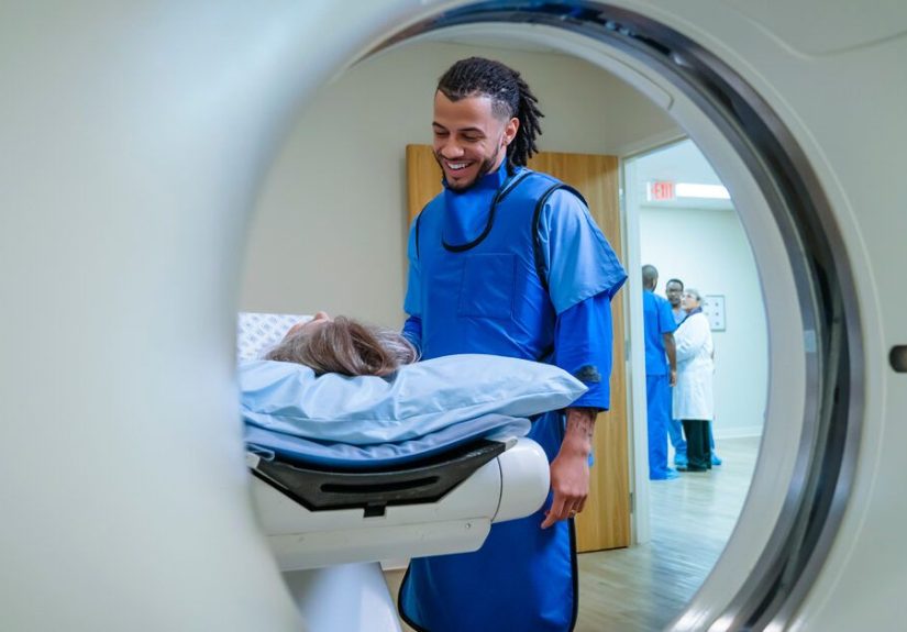
Step-by-step: what it’s like to get a CT or CTA
Before the scan
- You’ll be asked about contrast allergies, prior reactions, and kidney problems.
- You may be asked about medications and, in some cases, recent kidney function tests.
- Remove metal objects near the head/neck area when possible (hairpins, earrings, etc.).
During a non-contrast CT
- You lie on a table that slides into the scanner.
- Your job is to hold still (the scan is fastusually seconds to a few minutes).
- No needles are required for a standard non-contrast CT.
During a CTA
- An IV is placed for contrast injection.
- When contrast is injected, some people feel a brief warm flush or a metallic taste (temporary).
- Images are taken at specific timing so arteries “light up” clearly.
After the scan
- Most people can resume normal activity quickly unless they’re in emergency evaluation.
- You may be advised to hydrate afterward (especially if you received contrast), based on your clinician’s guidance.
- Results may be available quickly in emergency settings, but timing varies by facility and urgency.
How accurate is CT for aneurysm detection?
Accuracy depends on which CT test we’re talking about:
-
Non-contrast CT is primarily about detecting bleeding (like subarachnoid hemorrhage), not necessarily mapping
a small aneurysm directly. -
CTA is designed to detect aneurysms by showing detailed vessel anatomy, and it performs well for many clinically
significant aneurysmsespecially when image quality is high and the aneurysm is not extremely small.
Timing matters, too. For suspected SAH, non-contrast CT tends to be most sensitive earlier after symptom onset, and sensitivity
may decrease as time passes. That’s why clinicians sometimes add CTA, lumbar puncture, or other imaging if suspicion remains high
despite a negative initial CT.
Limitations and “false alarms” (because imaging is powerful, not magical)
Even excellent tests have limits. CTA may be less reliable when:
- The aneurysm is very small (tiny structures can be harder to distinguish from normal vessel curves).
- There’s motion during the scan (even slight movement can blur vessel edges).
- Nearby bone or calcification creates artifacts that complicate interpretation.
- Vessel anatomy is complex or there are multiple overlapping structures.
Sometimes a CTA finding is “indeterminate”meaning it’s not clearly normal or clearly an aneurysm.
In those cases, clinicians may recommend follow-up imaging (CTA or MRA) or proceed to DSA for definitive clarification.
Risks and safety: radiation, contrast, and the “should I worry?” question
Radiation exposure
CT uses X-rays, so there is radiation exposure. In emergencies, the benefit of quickly diagnosing a dangerous condition usually far outweighs
the theoretical long-term risk. Imaging teams also use protocols designed to keep doses as low as reasonably achievable while preserving image quality.
Contrast dye (CTA-specific)
Iodinated contrast is generally safe for most people, but it can cause side effects or reactions. Risks to discuss with your clinician include:
- Allergic-like reactions: ranging from mild (hives) to severe (rare).
- Kidney considerations: extra caution may be used in people with significantly reduced kidney function.
- Other medical factors: pregnancy or certain thyroid conditions may influence decisions.
If you’ve had a contrast reaction before, tell your care team clearly and early. There are established clinical strategies for risk assessment
and, when appropriate, premedication or alternative imaging approaches.
Smart questions to ask your clinician (so you don’t leave with “Wait, what?”)
- Am I getting a non-contrast CT, a CTA, or bothand why?
- If an aneurysm is found, what are its size, location, and features?
- Do I need follow-up imaging such as MRA or DSA?
- What symptoms should prompt me to seek emergency care?
- If contrast is used, what does my health history mean for contrast safety?
Bottom line
A standard head CT is often the fastest way to detect dangerous bleeding that can occur when an aneurysm ruptures.
A CT angiogram (CTA) goes a step further by highlighting blood vessels to help detect an aneurysm and define its anatomy.
Togetheroften alongside MRI/MRA or catheter angiography when neededthese tools help clinicians move from “something’s wrong” to
“here’s what it is, and here’s what we do next.”
Real-World Experiences: What People Often Report During CT/CTA for Aneurysm Evaluation (Extra Detail)
If you ask people what they remember most about getting scanned for a possible brain aneurysm, you might expect them to say,
“the machine” or “the images.” A lot of the time, they say something else: the waiting.
The scan itself can be surprisingly quick. The emotional partanticipation, uncertainty, and the mental replay of symptomscan feel much longer.
In emergency situations, many patients describe a fast-moving blur: triage questions, a neurologic exam,
then being rolled to CT. The non-contrast CT can feel almost anticlimactic: you lie down, the table moves, the scanner hums,
and before you’ve fully decided what face to make, it’s done. People often say the speed is comfortinglike the medical team
is taking the symptoms seriously and acting with purpose.
CTA has its own “signature moment.” Patients frequently mention the contrast injection: a brief warmth that can spread through the body
and a metallic taste that shows up uninvited, like a penny doing backflips on your tongue. It usually lasts seconds.
Many people also remember being told, “Hold still… hold your breath… okay, you can breathe.” It’s a small instruction with a big job:
motion can blur delicate vessel detail, and everyone wants the clearest pictures possible.
After imaging, experiences vary. In some ER settings, clinicians review results quickly and discuss next steps right away.
In other situations, people may be told that a radiologist will finalize the report and a specialist will interpret it in context.
That gap can feel stressful. A practical coping strategy many patients mention is asking one grounded question:
“What’s the plan while we wait?” Even if the plan is “monitor symptoms and keep you comfortable,” having a plan reduces the mental spiral.
For incidental findings (aneurysm found while scanning for something else), patients often report a different kind of shock:
not immediate fear from severe symptoms, but confusion“How can something in my brain be wrong if I feel okay?”
In these cases, follow-up conversations with specialists can be reassuring because decisions are often thoughtful and individualized:
watchful monitoring vs. treatment depends on aneurysm features, location, overall health, and risk profile.
People commonly say the most helpful moment was when someone explained the finding in plain English with a simple drawing:
“Here is the artery. Here is the bulge. Here’s what we’re watching for.”
Caregivers also carry a unique experience: they may not see the images, but they feel every minute of uncertainty.
Many caregivers describe relief when the team communicates clearlywhat the scan is checking, what a negative result means,
and what symptoms would change the urgency. Small communication details matter a lot, like being told whether the next step is CTA,
lumbar puncture, MRI/MRA, or specialist consultation. Clarity turns fear into a sequence of steps.
One more theme shows up again and again: people want to know what the result means for real life.
Not just “aneurysm yes/no,” but: “Can I drive? Can I work? Do I need follow-up? What should I do if I get another headache?”
If you’re in that position, it’s reasonable to ask for a short, written summary of the plan or the key pointsbecause your brain may be busy
doing other things (like worrying) during the conversation.
In the end, many patients describe CT/CTA as the start of a clearer story: turning scary, vague symptoms into concrete information.
Even when additional testing is needed, having imaging results can shift the experience from “unknown danger” to “understood problem,”
and that’s often the first real breath of relief.