Table of Contents >> Show >> Hide
- What a Shoulder MRI Can Show (and Why It’s Different from X-ray or Ultrasound)
- Purposes: When Doctors Typically Order a Shoulder MRI
- Types of Shoulder MRI: Standard MRI vs. MRI Arthrogram
- Procedure: What Happens During a Shoulder MRI (Step by Step)
- How to Prepare: Simple Tips That Actually Help
- Risks: What’s Real, What’s Rare, and What’s Mostly a Myth
- Results: What the Report Usually Includes
- FAQ: The Questions People Actually Ask
- Conclusion: The Big Takeaway
- Experiences: What It’s Like in Real Life (and What People Wish They’d Known) 500+ Words
Quick vibe check: A shoulder MRI is basically a super-detailed “photo shoot” for the soft tissues in your shouldertendons, ligaments, cartilage, labrum, muscles, and even subtle swelling. It uses a strong magnet and radio waves (not radiation), which is great news if the words “X-ray” and “extra exposure” make you suspicious.
That said, MRIs do have rules. The magnet is powerful enough that the MRI suite treats metal like it’s the main character. The goal of this guide is to explain why your clinician might order a shoulder MRI, what actually happens during the scan, and what risks (and non-risks) you should know aboutwithout making it sound like a sci-fi thriller.
What a Shoulder MRI Can Show (and Why It’s Different from X-ray or Ultrasound)
If your shoulder were a busy intersection, bones would be the roads and the soft tissues would be the traffic lights, signs, and everything that keeps the system from turning into chaos. X-rays are excellent at showing bones and joint alignment. MRIs shine when the question is, “What’s going on with the soft tissue?”
Common things a shoulder MRI helps evaluate
- Rotator cuff injuries (tendinopathy, partial tears, full-thickness tears)
- Labral tears (including SLAP-type injuries), often linked to instability or dislocations
- Biceps tendon issues (tendon irritation, tears, or related labral problems)
- Cartilage damage and early joint wear that may not show clearly on X-ray
- Bursitis and inflammatory changes around the joint
- Arthritis changes in soft tissue and bone marrow (plus related swelling)
- Infection or unusual fluid collections (in the right clinical context)
- Tumors or masses (rare, but MRI can help characterize tissue patterns)
There’s also a practical “why now?” reason: many clinicians start with an X-ray for shoulder pain to screen for obvious bone or alignment issues, then escalate to ultrasound or MRI depending on the suspected problem. Ultrasound can be very effective for rotator cuff evaluation (especially in experienced hands), while MRI is often preferred when the concern is intra-articular problemslike labral tearsor when multiple structures may be involved.
Purposes: When Doctors Typically Order a Shoulder MRI
A shoulder MRI isn’t usually the first stop for every ache. It’s often ordered when symptoms, exam findings, or earlier imaging suggest something more specific than “general soreness.” Here are the common situations where a shoulder MRI earns its paycheck.
1) Persistent pain that doesn’t match the “simple strain” story
If you’ve rested, tried activity changes, maybe done physical therapy, and the shoulder still acts like it’s holding a grudge, an MRI can clarify whether something structural is keeping the problem alivelike a tear, significant inflammation, or cartilage damage.
2) Suspected rotator cuff tear (especially after injury)
A sudden pop, weakness lifting the arm, pain at night, or loss of function after a fall can raise suspicion for a tear. MRI can help determine the size and location of a tear, which can influence treatment planning.
3) Instability, dislocations, or “it feels like it slips”
Recurrent dislocations or a shoulder that feels unstable can be associated with labral injuries. MRI (and sometimes MRI arthrogrammore on that soon) can help identify labral damage and related structural issues.
4) Sports-related or repetitive overhead stress
Throwing athletes, swimmers, volleyball players, painters, mechanicsanyone who spends a lot of time overheadcan develop a mix of tendon irritation, bursitis, impingement patterns, and labral wear. MRI can help sort out the “which structure is actually complaining?” question.
5) Surgical planning or “Why didn’t the first plan work?”
Sometimes MRI is used to guide decisions about surgery vs. non-surgical treatment, or to evaluate ongoing symptoms after a prior repair (keeping in mind that post-surgical shoulders can look different on imaging).
Types of Shoulder MRI: Standard MRI vs. MRI Arthrogram
Not all shoulder MRIs are the same flavor.
Standard shoulder MRI (no joint injection)
This is the most common type. You lie still, the scanner does its thing, and images are captured in multiple planes. A standard MRI may be done with or without IV contrast depending on the question.
MRI arthrogram (contrast injected into the joint)
If your clinician strongly suspects a labral tear or subtle intra-articular issue, they may recommend an MRI arthrogram. This involves an injection of contrast material directly into the shoulder joint (typically done under imaging guidance) before the MRI images are taken. The idea is simple: the contrast helps outline the joint structures, which can make certain tears easier to spot.
Translation: Standard MRI is great for many problems. MRI arthrogram can be especially useful when the labrum is the prime suspect and the case needs a brighter flashlight.
Procedure: What Happens During a Shoulder MRI (Step by Step)
If you’ve never had an MRI, the unknown is usually worse than the scan itself. Here’s the usual flow in everyday terms.
Step 1: Safety screening (a.k.a. the “metal interview”)
You’ll answer questions about implants, prior surgeries, metal fragments (especially in the eyes), devices, and certain medical conditions. This is not paperwork theaterthe magnet is the reason. Many modern implants are MRI-conditional, but the facility needs details.
Step 2: Changing and removing metal items
You’ll be asked to remove jewelry, watches, hair pins, certain clothing with metal (zippers, snaps), and anything else metallic. You may change into a gown. Cards and electronics don’t love MRI magnets either, so they’re usually kept out of the scanning area.
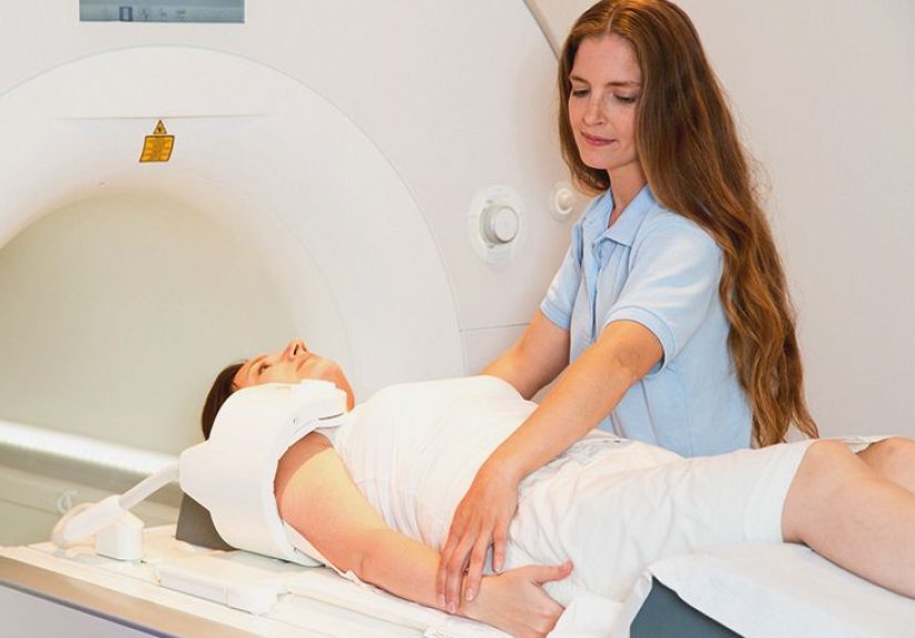
Step 3: Positioning (the “please don’t move” era begins)
You’ll lie on the table, typically on your back. The technologist positions your arm and shoulder and places a special device called a coil around the shoulder. The coil helps the scanner capture clearer images.
Step 4: The scan itself
The table slides into the MRI machine, which is often tunnel-shaped (though some facilities use wider-bore or open designs). The scanner is noisythink rhythmic thumping and tapping like a very committed woodpecker with a music career. Earplugs or headphones are commonly offered.
Most shoulder MRI exams take roughly 30 to 60 minutes, depending on the protocol and whether contrast is used. The key job for you is to stay as still as possible, because motion can blur images.
Step 5: If contrast is needed
If your MRI includes contrast, it may be:
- IV gadolinium-based contrast (in a vein), often used when the goal is to evaluate inflammation, infection, tumors, or certain complex questions
- Intra-articular contrast (MRI arthrogram), injected into the joint before imaging to highlight the labrum and joint surfaces
Step 6: After the scan
Most people can return to normal activities right away. If you received a sedative for anxiety or claustrophobia, you may need someone to drive you home and you may be advised to take it easy for the rest of the day.
How to Prepare: Simple Tips That Actually Help
Before you arrive
- Bring implant/device info if you have it (cards for pacemakers, surgical records, device brand/model details).
- Tell your clinician if you’re pregnant, have kidney disease, or have had reactions to contrast in the past.
- Ask about anxiety/claustrophobia options in advance if enclosed spaces make your brain hit the panic button.
What to wear
Comfortable clothes without metal. If you’re not sure, the facility will often provide a gown anyway.
Food and meds
Most shoulder MRIs do not require fasting. The main exception is if you’ll receive sedation or if the facility has specific instructionsfollow what your imaging center tells you.
Risks: What’s Real, What’s Rare, and What’s Mostly a Myth
Here’s the reassuring headline: MRI does not use ionizing radiation. That’s one major risk category you can cross off. The meaningful risks relate to (1) the magnet + metal and (2) contrast in select cases.
1) Metal and implanted devices (the biggest safety issue)
The MRI magnet is strong. Certain implants or metal fragments can pose risks or affect image quality. That’s why screening is strict. Many devices are safe under specific conditions, but the imaging team needs to confirm compatibility.
2) Contrast risks (uncommon, but worth knowing)
If your exam uses a gadolinium-based contrast agent (GBCA), side effects are usually mild when they occur. A small number of people may have nausea, headache, or a mild allergic-type reaction. Severe allergic reactions are rare.
Kidney disease and NSF: The most serious known contrast-related complication is nephrogenic systemic fibrosis (NSF), which is rare and occurs mainly in people with severe kidney impairment. This is why imaging teams may check kidney function before giving contrast in higher-risk patients.
Gadolinium retention: Small amounts of gadolinium can be retained in the body after some contrast MRIs. U.S. regulators have noted that, to date, the only clearly established harm associated with retention is NSF in a small subgroup of patients with serious kidney failure. Research continues, and clinicians weigh risks vs. benefits when deciding on contrast.
3) Claustrophobia and anxiety (common, manageable)
Some people feel uneasy in the scanner. The good news: facilities commonly offer ear protection, communication with the technologist, and sometimes music. In certain cases, mild sedation may be used.
4) Noise and minor discomfort
The machine is loud and the shoulder position can feel awkwardespecially if you’re already sore. The scan is painless, but holding still in one position can be uncomfortable. If something hurts, tell the technologist before the scan begins so they can adjust positioning when possible.
5) Pregnancy considerations
MRI is sometimes used during pregnancy when clinically necessary, but contrast is generally approached more cautiously. If there’s any chance you’re pregnant, tell your care team so they can choose the safest approach for your situation.
Results: What the Report Usually Includes
Your MRI images are interpreted by a radiologist, who creates a report for your ordering clinician. Reports often describe:
- Rotator cuff tendons (inflammation vs. tear, location, extent)
- Labrum (signs of tearing or detachment)
- Biceps tendon and anchor area
- Cartilage and joint surfaces
- Bursa and signs of impingement-related inflammation
- Bone marrow changes (stress reactions, bruising, degenerative changes)
- Fluid collections or signs of infection (if suspected clinically)
One important reality check: Imaging findings should match symptoms and exam findings. MRI can show changes that don’t cause pain (especially in athletes or as we age). The best care decisions come from combining the report with your real-life symptoms and functional limitations.
FAQ: The Questions People Actually Ask
“Will a shoulder MRI show a rotator cuff tear?”
Often, yes. MRI can evaluate rotator cuff tendons in detail, including partial and full tears, tendon inflammation, and related muscle changes.
“Is an MRI arthrogram better for labral tears?”
In many cases, it can be. Injecting contrast into the joint may help outline the labrum and subtle intra-articular tears more clearly. Your clinician chooses this when the clinical question calls for it.
“What if I have a pacemaker or implants?”
Don’t assume it’s an automatic “no,” but do assume it requires careful review. Many devices can be scanned under specific protocols. Bring device details so the imaging team can confirm safety conditions.
“How long does a shoulder MRI take?”
Commonly 30–60 minutes, depending on the protocol and whether contrast is used.
“What are alternatives if MRI isn’t possible?”
Depending on the question, options may include ultrasound (especially for rotator cuff evaluation), CT, CT arthrogram, or X-ray for bone-related concerns. The “best” alternative depends on what your clinician is trying to confirm or rule out.
Conclusion: The Big Takeaway
A shoulder MRI is one of the best tools for seeing what’s happening in the soft tissues that make the shoulder workespecially when the issue may involve the rotator cuff, labrum, cartilage, or hidden inflammation. The procedure is noninvasive, doesn’t use radiation, and is generally very safe when proper screening is done. The main risk revolves around metal safety and, in select cases, contrast use (particularly in severe kidney disease).
If you’re scheduled for a shoulder MRI, the most helpful things you can do are: provide accurate implant history, follow the imaging center’s prep instructions, and tell the team about anxiety, kidney disease, pregnancy, or past contrast reactions. And remember: the MRI report is a powerful cluenot the whole mystery novel. Your symptoms and exam still matter.
Experiences: What It’s Like in Real Life (and What People Wish They’d Known) 500+ Words
Even when someone understands the “science” of an MRI, the real-life experience can still feel weirdly emotionalbecause it’s your body, your pain, and a machine that sounds like it’s building a spaceship in the next room. People often describe the day of the scan as a mix of “This is probably fine” and “Why is my shoulder suddenly more dramatic than ever?”
The appointment check-in is usually the calmest part. You answer safety questions, change clothes (goodbye, hoodie zipper), and meet a technologist who has clearly explained the MRI process 4,000 times. A lot of people feel reassured right here because the staff tends to be practical and direct: what to remove, how to position, how long it might take, and how to communicate during the scan.
Positioning can be the most uncomfortable moment. If your shoulder already hurts, lying still with the arm placed “just so” can feel like an endurance test. Many people say they wish they’d spoken up earlier if the position felt unsustainable. Technologists can often adjust supports, add padding, or tweak your arm angle before the scan starts. The goal is not to win an award for toughness; the goal is clear images without you suffering.
The sound surprises almost everyone. MRI noises aren’t just loudthey’re patterned and intense, like industrial techno created entirely by magnets. Some people find the rhythm oddly calming. Others think, “So this is how a toaster would sound if it had unresolved anger issues.” Earplugs help a lot, and some facilities offer headphones or music. People who do best tend to treat the noise like weather: it’s happening, it’s not personal, and it will pass.
Claustrophobia is more common than people admit. Many patients report that they felt totally fine… until the table moved into the scanner, and then their brain suddenly ran a full emergency simulation. What helps in those moments? Knowing you can talk to the technologist, focusing on slow breathing, keeping your eyes closed (for some), and remembering the scan is broken into short sequences. For people who are anxious about enclosed spaces, scheduling the scan at a place with a wider-bore MRI or discussing calming options ahead of time can make the experience dramatically easier.
Contrast can add a layer of “Wait, what now?” If you need IV contrast, many people barely notice beyond the quick IV placement. If you need an arthrogram injection into the shoulder joint, patients often describe it as more mentally intimidating than physically painfulbecause it’s a joint injection, and joints have a reputation. The reality is that the process is typically done with guidance to improve accuracy, and while it can be uncomfortable, it’s usually brief. People often say the bigger challenge is the waiting afterward: “Did it work? Did they get the right spot? Is it supposed to feel tight?” (Those are normal thoughts. Ask your care team what sensations are expected.)
The hardest part is sometimes the waiting for results. Many patients describe the gap between scan and interpretation as the most stressful phase. They’re replaying every shoulder movement in their head like a detectiveexcept the detective is also the suspect. If you’re in that window, it can help to remember: MRI findings need context. Some “abnormalities” are common, especially in active people, and not every tear equals surgery. The report is a tool your clinician uses to match images with symptoms and build a sensible plan.
Afterward, most people feel reliefeven if nothing is “fixed” yet. There’s something comforting about having more clarity. Whether the MRI shows a clear cause (like a tear) or rules out major problems, the scan often turns the shoulder story from “mystery pain” into “here’s what we’re dealing with,” which is a better place to start recovery.