Table of Contents >> Show >> Hide
- Interventional Radiology, Explained Like a Normal Human Being
- How Interventional Radiology Works
- What Conditions Can Interventional Radiology Treat?
- Common Interventional Radiology Procedures
- Why Doctors and Patients Choose IR
- Who Performs Interventional Radiology?
- What to Expect as a Patient
- The Future of Interventional Radiology
- Final Thoughts
- Real-World Experiences With Interventional Radiology
Interventional radiology sounds like one of those phrases people nod at politely while secretly thinking, “Right, and what exactly does that mean?” Fair question. The short version is this: interventional radiology, often called IR, is a medical specialty that uses imaging to guide tiny tools through the body to diagnose or treat disease with as little disruption as possible. Think of it as precision medicine with a map, a camera, and a very steady hand.
Instead of making large incisions, an interventional radiologist often works through a pinhole opening in the skin using catheters, needles, wires, balloons, stents, or other miniature devices. The doctor watches the path in real time with imaging such as X-ray fluoroscopy, CT, ultrasound, or MRI. The goal is simple but powerful: treat the problem directly while causing less pain, fewer complications, and a shorter recovery than traditional open surgery in many cases.
That is why interventional radiology procedures have become a big deal in modern medicine. They are now used across cancer care, vascular disease, women’s health, liver disease, kidney problems, dialysis access, pain treatment, and much more. In other words, IR is not a niche side character anymore. It is very much in the main cast.
Interventional Radiology, Explained Like a Normal Human Being
If surgery is the classic front-door entrance, interventional radiology is the “quiet side entrance with a keycard and a detailed floor plan.” Rather than opening up a large section of the body, IR doctors use image-guided treatment to reach the exact target through a very small access point. That target might be a blocked artery, a bleeding blood vessel, a tumor, a painful fracture, a fibroid, a clot, or a narrowed duct.
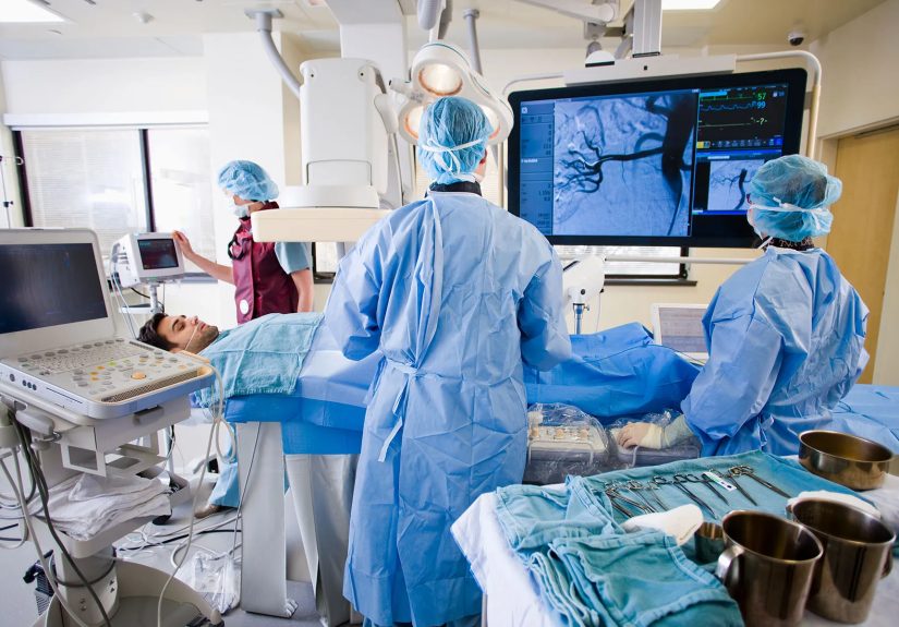
The imaging is what makes the specialty work. It is not just about seeing the body; it is about navigating inside it with extraordinary accuracy. A doctor can guide a catheter into a liver artery, place a stent in a narrowed vessel, sample tissue from a suspicious mass, drain an infection, or destroy a tumor with heat or cold. The body is still complicated, of course. IR just gives medicine a much better GPS.
How Interventional Radiology Works
The Imaging Tools Behind the Magic
Interventional radiologists rely on several imaging methods, depending on the procedure and the body part involved:
Fluoroscopy: a live X-ray that helps doctors watch wires, contrast dye, and catheters move through blood vessels and organs.
Ultrasound: useful for guiding needles into soft tissues, blood vessels, fluid collections, and certain tumors without using radiation.
CT: excellent for detailed cross-sectional guidance, especially for biopsies, drainage procedures, and some ablations.
MRI: less common in day-to-day IR than ultrasound or fluoroscopy, but valuable in selected cases where soft-tissue detail really matters.
These tools help the doctor work with remarkable precision. That precision is one reason minimally invasive procedures can often replace or complement surgery.
What the Doctor Actually Uses
The equipment is tiny compared with what most people imagine. A procedure may involve a needle, a guidewire, a slim catheter, a balloon to open a narrowed vessel, a metal mesh stent to keep it open, particles to block blood flow, or a probe that destroys tumor tissue with heat, cold, or energy. It is highly technical, but the basic idea is elegant: go directly to the problem and fix it with the least collateral drama possible.
What Conditions Can Interventional Radiology Treat?
One of the most surprising things about IR is how many body systems it touches. This is not a one-trick specialty. It is more like a toolkit used across medicine.
Blood Vessel Problems
IR is widely used for vascular disease. That includes angioplasty and stenting for narrowed arteries, clot removal for deep vein thrombosis or pulmonary embolism in selected cases, embolization to stop bleeding, and treatment of aneurysms or abnormal blood vessels. For some patients, these treatments can restore blood flow, reduce stroke risk, prevent limb complications, or control life-threatening bleeding without a major operation.
Cancer Care
Interventional oncology is one of the fastest-growing areas in IR. Doctors can perform biopsies to diagnose tumors, then use targeted therapies such as ablation, chemoembolization, and radioembolization to treat certain cancers, especially in organs like the liver, kidney, lung, and bone. These treatments can destroy tumors directly or reduce their blood supply while preserving more healthy tissue than broader approaches. In some cases, IR supports surgery, chemotherapy, radiation, or transplant care rather than replacing them.
Women’s and Men’s Health
IR also treats conditions that affect daily quality of life in a big way. Uterine fibroid embolization can shrink fibroids by blocking their blood supply. Prostate artery embolization can help some men with symptoms from an enlarged prostate. Pelvic congestion syndrome, varicoceles, and certain fertility-related or pain-related vascular issues may also be managed with image-guided techniques.
Liver, Kidney, and Digestive Conditions
Interventional radiologists place biliary drains, open narrowed ducts, create shunts such as TIPS for portal hypertension, sample liver tissue, and treat kidney or liver tumors with ablation or embolization. They also place feeding tubes and manage fluid collections or abscesses that need drainage.
Biopsy, Drainage, Access, and Pain Procedures
Some of the most common IR procedures are not glamorous, but they are incredibly useful. Image-guided biopsy helps doctors diagnose disease without open surgery. Drainage procedures can relieve infections or fluid buildup. IR physicians place ports, PICC lines, dialysis access devices, and IVC filters when appropriate. They may also treat painful vertebral compression fractures with vertebroplasty or kyphoplasty in selected patients.
Common Interventional Radiology Procedures
Angioplasty and stenting: opening narrowed blood vessels to improve circulation.
Embolization: blocking blood flow on purpose to stop bleeding, shrink fibroids, reduce tumor blood supply, or treat abnormal vessels.
Ablation: destroying tumors or painful lesions with heat, cold, or electrical energy.
Biopsy: removing a small tissue sample with imaging guidance for diagnosis.
Drain placement: removing infected fluid or relieving pressure from blocked systems.
Venous access placement: inserting ports, PICCs, or dialysis-related devices.
Thrombectomy or thrombolysis: removing or dissolving clots in carefully selected cases.
Why Doctors and Patients Choose IR
The Biggest Benefits
The strongest selling point of interventional radiology is that it often offers a less invasive path to the same destination. That can mean:
Smaller incisions or punctures instead of large surgical cuts.
Less pain and, in many cases, less need for heavy anesthesia.
Lower risk of some complications compared with open surgery.
Shorter hospital stays or even same-day treatment.
Faster return to normal life, which is always popular with people who have laundry, jobs, and a very opinionated dog waiting at home.
For medically complex patients, these advantages matter even more. Someone who is older, frail, or dealing with multiple health conditions may tolerate a minimally invasive option better than a traditional operation.
The Important Reality Check
IR is impressive, but it is not magical and it is not always the best choice. Some conditions still require surgery. Some patients need a combination of treatments. Certain procedures involve contrast dye, radiation exposure, bleeding risk, infection risk, or the possibility that the first treatment will not fully solve the problem. The best use of IR comes from good patient selection and close teamwork among radiologists, surgeons, oncologists, cardiologists, hepatologists, nephrologists, and primary care clinicians.
Who Performs Interventional Radiology?
An interventional radiologist is not just someone who reads scans in a dark room and occasionally emerges like a wizard from a cave. These are board-certified physicians with extensive training in diagnostic imaging, image-guided procedures, and patient management before, during, and after treatment.
Modern IR training includes medical school, internship, diagnostic radiology training, and dedicated interventional radiology residency or fellowship training. The specialty also involves clinic visits, consultation, periprocedural care, and follow-up. In other words, this is not just “doing procedures.” It is a full clinical specialty.
What to Expect as a Patient
Before the Procedure
You usually start with a consultation, imaging review, lab work if needed, and instructions about medications, food, and transportation. Some blood thinners may need special planning. Some procedures require fasting. Others are simpler and move quickly.
During the Procedure
Many IR treatments use local numbing medicine plus moderate sedation, though some need deeper anesthesia and some need very little at all. The team cleans the skin, uses imaging to guide the instruments, and performs the treatment through a tiny opening. Patients often feel pressure rather than sharp pain, but the exact experience depends on the procedure.
After the Procedure
Recovery may be brief, especially for outpatient procedures. Some patients go home the same day. Others stay overnight for monitoring, pain control, or observation. You may receive instructions about activity limits, wound care, hydration, or follow-up imaging. Recovery tends to be easier than many people expect, which is one reason IR earns so many “Wait, that was it?” reactions.
The Future of Interventional Radiology
IR keeps expanding because imaging keeps getting better and devices keep getting smarter. New technologies in navigation, robotics, artificial intelligence, and tumor targeting are pushing the field forward. Cancer care is a major area of growth, especially in therapies that can destroy tumors with precision while sparing nearby tissue. There is also ongoing work to improve access, reduce radiation dose, increase safety, and bring image-guided treatments to patients who previously had limited options.
That future matters because healthcare is steadily moving toward treatments that are more targeted, less disruptive, and more personalized. Interventional radiology fits that direction almost suspiciously well.
Final Thoughts
So, what is interventional radiology? It is the medical specialty that blends imaging, engineering, and hands-on treatment to solve problems inside the body through the smallest practical opening. It can diagnose disease, stop bleeding, open blocked vessels, destroy tumors, shrink fibroids, drain infections, support dialysis, relieve pain, and spare many patients from larger operations.
Its appeal is not hard to understand. When medicine can treat the right target with more precision, less pain, and less recovery time, that is a win for patients and clinicians alike. Interventional radiology is not the answer to every medical question, but for the right condition and the right patient, it can be one of the smartest answers in the room.
Real-World Experiences With Interventional Radiology
If you ask patients what interventional radiology feels like in real life, the most common theme is surprise. Not surprise that the technology exists, but surprise that something with such a dramatic name can feel so controlled, calm, and efficient. Many people arrive expecting a full surgical production. Instead, they find a carefully choreographed procedure, a focused team, a lot of monitors, and a process that is often faster and less overwhelming than they imagined.
A typical patient experience starts with uncertainty. Someone may have been told they have a liver tumor, painful uterine fibroids, a blocked artery, a fluid collection, or a clot. They are worried, naturally, and then they hear there may be a treatment that does not require a large incision. That first IR consultation often changes the tone of the conversation. The doctor explains the anatomy, shows the imaging, outlines the goal, and describes how a catheter, needle, or probe can be guided precisely to the problem area. For many patients, that is the moment the situation starts to feel manageable.
On procedure day, the experience is usually more “high-tech calm” than “medical chaos.” Patients are checked in, reviewed by nurses, and prepped with an IV. In the procedure suite, they may notice large imaging screens, bright lights, and a lot of equipment that looks intimidating until the team starts explaining what everything does. The staff in interventional radiology tends to be very procedure-focused and detail-oriented, which patients often find reassuring. There is comfort in watching people move like they have done this a thousand times, because they usually have.
During the procedure itself, many patients are surprised by how little they feel. They may be sleepy from sedation, aware enough to hear instructions, but comfortable enough to remember only bits and pieces. Some describe feeling pressure, warmth from contrast dye, or brief discomfort when the doctor reaches the treatment site, but not the kind of pain they associated with “having something done.” In biopsy or drain placement cases, the experience can feel almost anticlimactic. In embolization or ablation cases, there may be more recovery afterward, but still far less than many expected when they first heard the words “tumor treatment” or “vascular procedure.”
The recovery experience is another part that stands out. Some patients rest for a few hours and go home the same day. Others stay overnight, especially after more involved treatments. What they often remember most is the contrast between the seriousness of the condition and the relative simplicity of the recovery. Instead of a long incision, they may have a tiny bandage. Instead of days in the hospital, they may be back in their own bed that night. Instead of a long delay before normal life resumes, they may be walking, eating, and functioning much sooner than they thought possible.
Families notice it too. They expect surgery-level disruption and instead see something more targeted and contained. That does not make IR trivial. These are still real medical procedures with real risks and real follow-up. But the experience often feels more precise, more modern, and more patient-friendly. For many people, interventional radiology is the first time they realize that treatment does not always have to be bigger to be better. Sometimes the smartest medicine is the medicine that knows how to do more while bothering the body less.