Table of Contents >> Show >> Hide
- Why Early Detection Matters So Much
- Screening vs. Detection vs. Diagnosis: What’s the Difference?
- Who Should Get Screened, and When?
- Screening Tools Doctors Use to Find Breast Cancer
- What Happens If Your Screening Mammogram Is Abnormal?
- How Doctors Confirm a Breast Cancer Diagnosis
- Staging: Understanding How Advanced the Cancer Is
- Talking with Your Doctor: Questions That Help
- Common Myths About Breast Cancer Screening
- Real-Life Experiences: What Screening & Diagnosis Can Feel Like
- Bottom Line: Screening Is a Tool, Not a Verdict
Hearing the words “breast cancer screening” can make anyone’s shoulders tense up a little.
The machines look intense, the medical jargon sounds like a foreign language, and trying to
figure out when to start mammograms can feel like planning a rocket launch. The good news?
Doctors have a clear system for finding breast cancer as early as possible, and most of it
is far less scary once you know what actually happens.
This guide walks you through how breast cancer is detected, when screening typically starts,
which tests are used, and what happens if something suspicious shows up. Think of it as a
friendly roadmap to a process that’s designed to protect your health, not terrify you.
Why Early Detection Matters So Much
Breast cancer is common, but the timing of when it’s found can make a huge difference.
Cancers caught early are more likely to be smaller, have not spread to lymph nodes or distant
organs, and are often easier to treat with less aggressive therapies. Decades of research show
that regular screening mammograms help reduce deaths from breast cancer, because they can find
tumors before they cause symptoms like lumps, pain, or skin changes.
In simple terms: screening is about finding trouble early, when doctors still have many good
options. Waiting until you “feel something” can mean the cancer has already had time to grow or spread.
Screening vs. Detection vs. Diagnosis: What’s the Difference?
These three terms are related, but they don’t mean the same thing:
-
Screening is checking people who feel completely fine, to see if there are
any hidden early signs of cancer. Mammograms are the main breast cancer screening test. -
Detection is the moment something abnormal is found maybe a spot on a
mammogram or a lump in the breast. -
Diagnosis is confirming what that abnormality actually is, usually by
doing a biopsy and having a pathologist examine cells under a microscope.
You can think of screening as the “smoke detector,” detection as “spotting the smoke,” and
diagnosis as “figuring out whether it’s steam from your shower or an actual fire.”
Who Should Get Screened, and When?
There’s no single global rule for breast cancer screening, but major U.S. organizations now
agree on some key ideas, especially for people at average risk (no strong
family history, no known high-risk gene mutation, and no prior chest radiation at a young age).
Guidelines for People at Average Risk
The U.S. Preventive Services Task Force (USPSTF) currently recommends that women and others
assigned female at birth who are at average risk get a screening mammogram every two years
starting at age 40 and continuing through age 74. This “every other year” schedule (called
biennial screening) is meant to balance the benefits of catching cancers early with
the downsides of false alarms and extra testing.
The American Cancer Society (ACS) offers slightly different age and timing guidance:
- Ages 40–44: You have the option to start yearly screening if you choose.
- Ages 45–54: Yearly mammograms are recommended.
- Age 55 and older: You can switch to mammograms every two years or continue yearly,
as long as you’re in good health and expected to live at least 10 more years.
Different professional groups may vary a bit, but they all emphasize shared decision-making
talking with your doctor about your personal risk factors, preferences, and comfort level with
more frequent testing.
What About Screening After Age 75?
For people older than 74, the evidence isn’t as clear, and major guidelines say there isn’t
enough data to make a one-size-fits-all recommendation. In real life, many clinicians look at
overall health, other medical issues, and life expectancy. Someone who’s 78, active, and
generally healthy may choose to keep getting mammograms, while someone who is more frail or
dealing with serious illnesses may reasonably decide to stop.
Who Is Considered High Risk?
Some people have a higher-than-average risk of breast cancer and may need earlier or more
intensive screening. You may be considered high risk if you:
- Have a strong family history of breast or ovarian cancer (especially in close relatives at younger ages).
-
Carry certain inherited genetic mutations (like BRCA1 or BRCA2) or have
a strong suspicion based on family patterns. - Had radiation therapy to the chest before age 30, for example to treat lymphoma.
- Have certain high-risk breast conditions found on biopsy.
For these individuals, doctors often recommend both yearly mammograms and yearly breast MRI,
sometimes starting as early as age 30, though the exact plan depends on your personal situation.
Screening Tools Doctors Use to Find Breast Cancer
Breast cancer screening is not one single test. Doctors have a toolkit, and they choose pieces
based on your risk, age, breast tissue, and any symptoms you might have.
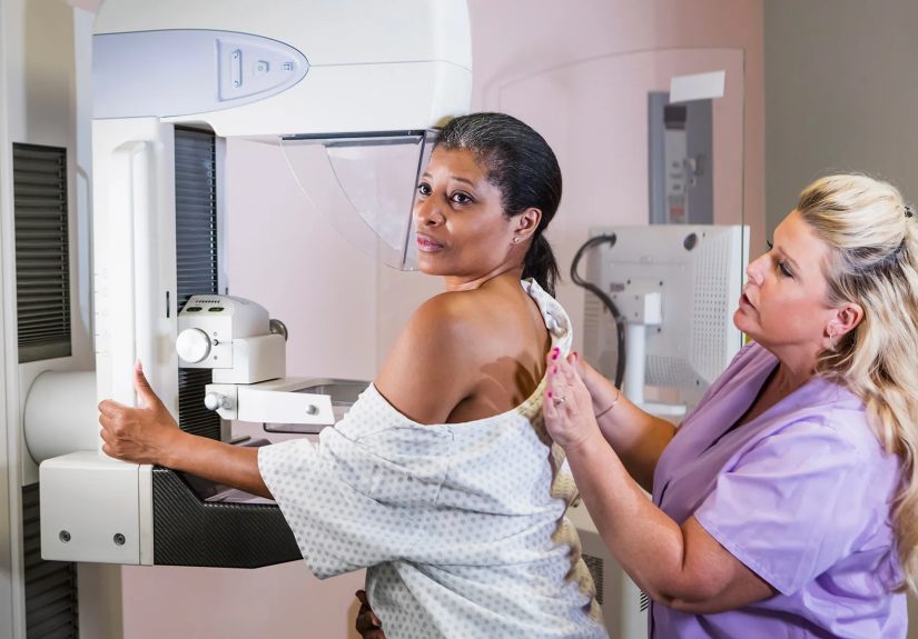
Mammograms: The Workhorse of Screening
A mammogram is a low-dose X-ray of the breast. It can show tiny calcifications,
distortions, or masses long before they can be felt. Modern mammography comes in two main flavors:
- Digital mammography (2D) – Traditional images taken from two views of each breast.
-
Digital breast tomosynthesis (3D mammography) – Takes multiple images from
different angles that a computer reconstructs into thin “slices,” making it easier to see through
overlapping tissue, especially in dense breasts.
The actual exam usually takes about 20 minutes. Yes, the compression is uncomfortable (no one is
writing love poems about it), but it lasts only seconds for each image and dramatically improves
picture quality and lowers radiation.
Clinical Breast Exams and Breast Self-Awareness
A clinical breast exam is when a health care professional carefully examines your
breasts and underarm areas for lumps, thickening, or changes in skin or nipples. Many major
organizations do not recommend clinical breast exams alone as a primary screening method anymore
for people at average risk, but they are still important for evaluating symptoms and for people
who notice changes between mammograms.
Instead of rigid “self-exam” routines, experts now often talk about breast self-awareness
simply being familiar with how your breasts usually look and feel, and letting your provider know
if something seems off.
Ultrasound: Helpful in Dense Breasts or When Something Looks Unclear
A breast ultrasound uses sound waves (no radiation) to create images. It’s often
used to look more closely at an area that appeared suspicious or uncertain on a mammogram.
Ultrasound can help tell whether a lump is a solid mass or a fluid-filled cyst and is especially
useful in people with dense breast tissue.
Breast MRI: Powerful Imaging for Specific Situations
Breast MRI (magnetic resonance imaging) uses a powerful magnet, radio waves,
and a computer to create highly detailed pictures. It’s more sensitive than mammography, which
can be a big advantage in high-risk patients, but that sensitivity also means it can pick up
more non-cancer findings that still need follow-up.
Breast MRI is usually reserved for:
- Screening people at high risk of breast cancer (often in addition to mammograms).
- Evaluating the extent of cancer once it’s been diagnosed.
- Clarifying tricky or inconclusive findings from other imaging tests.
What Happens If Your Screening Mammogram Is Abnormal?
First things first: an abnormal screening mammogram does not automatically mean
you have breast cancer. Most callbacks turn out to be benign (non-cancerous) findings or normal
tissue that simply looked unusual on the first set of images.
If something looks suspicious or unclear, the radiology team may:
- Take additional mammogram views (diagnostic mammogram) focused on the area of concern.
- Perform a targeted ultrasound to get more detail.
- Recommend a breast MRI if needed.
After these extra images, the radiologist will decide whether:
- The area looks normal and you can go back to routine screening.
- The finding is probably benign but should be watched with a short-interval follow-up (for example, a repeat image in six months).
- The area is suspicious enough that a biopsy is recommended.
How Doctors Confirm a Breast Cancer Diagnosis
Imaging can tell doctors where a suspicious area is and what it might look like, but it cannot
prove whether it’s cancer. For that, they need tissue. That’s where a biopsy comes in.
Common Types of Breast Biopsies
Your care team chooses the type of biopsy based on where the abnormal area is and how it appears
on imaging:
-
Core needle biopsy: A hollow needle removes small cylinders (“cores”) of tissue,
usually guided by ultrasound, mammography (stereotactic biopsy), or MRI. -
Vacuum-assisted biopsy: A special device uses gentle suction to collect more
tissue through a single small incision. -
Fine-needle aspiration (FNA): A very thin needle removes cells or fluid; this
is sometimes used for lumps or cysts. -
Surgical (excisional) biopsy: A surgeon removes all or part of a suspicious
area in the operating room, usually when needle biopsy is not possible or has not given a
clear answer.
The tissue goes to a pathologist, a doctor trained to identify disease under the
microscope. The pathology report is the official word on whether cancer is present and, if so,
what type.
What the Pathology Report Tells You
If breast cancer is found, the report will include details such as:
- Type of cancer (for example, invasive ductal carcinoma or invasive lobular carcinoma).
- Whether it is invasive (has spread beyond the ducts or lobules) or non-invasive (like DCIS).
- Tumor grade (how abnormal the cells look and how fast they appear to be growing).
-
Hormone receptor status (whether the cells have estrogen and/or progesterone receptors, which
may respond to hormone-blocking treatments). - HER2 status, a protein that can influence growth and treatment options.
All of this information helps the oncology team build a tailored treatment plan.
Staging: Understanding How Advanced the Cancer Is
After diagnosis, doctors determine the stage of breast cancer. Staging is a way
of describing how big the tumor is, whether lymph nodes are involved, and whether the cancer has
spread to other parts of the body.
Most staging systems use the TNM framework:
- T (Tumor): Size and extent of the main tumor in the breast.
- N (Nodes): Whether cancer is in nearby lymph nodes (especially under the arm).
- M (Metastasis): Whether cancer has spread to distant organs like bone, liver, or lungs.
These details, along with tumor grade and key biomarkers, are combined into an overall stage group:
- Stage 0: Non-invasive disease (like ductal carcinoma in situ, or DCIS).
- Stage I: Small, early-stage invasive cancers with limited or no lymph node involvement.
- Stage II–III: Larger tumors and/or more lymph node involvement, but still potentially curable.
- Stage IV: Cancer that has spread to distant sites (metastatic breast cancer).
While the word “stage” can sound intimidating, its main purpose is practical: to guide treatment
and help estimate prognosis, not to define you as a person.
Talking with Your Doctor: Questions That Help
Medical appointments can go by quickly, and it’s easy to walk out thinking, “I wish I’d asked…”.
Consider bringing a written list of questions, such as:
- Based on my age and history, when should I start or continue mammograms?
- Am I considered average risk or high risk for breast cancer?
- Should I have additional screening, like ultrasound or breast MRI?
- What did my mammogram or other imaging actually show?
- Why are you recommending (or not recommending) a biopsy?
- If cancer is found, what stage is it and what treatment options are available?
Bringing a friend or family member, asking for written summaries, or using a patient portal to
review notes afterward can help you process everything more calmly.
Common Myths About Breast Cancer Screening
“If I Don’t Feel a Lump, I Don’t Need a Mammogram”
Unfortunately, many early breast cancers don’t cause pain or obvious symptoms. Mammograms can
detect changes that are too small to feel. Waiting to schedule screening until you notice
something may mean finding cancer at a later stage.
“Mammograms Expose Me to Too Much Radiation”
Mammograms use low-dose X-rays. The amount of radiation is small and considered safe for regular
screening, especially compared with the very real benefit of catching cancer early. The
risk-benefit balance strongly favors screening for most people in recommended age groups.
“If I Get Called Back, I Probably Have Cancer”
Callback rates vary, but most people who return for extra views or ultrasound do not
have cancer. Many times, it’s simply dense tissue overlapped in a funny way, or a benign cyst,
or something that just needs a closer look for reassurance.
Real-Life Experiences: What Screening & Diagnosis Can Feel Like
Guidelines and acronyms are helpful, but they don’t fully capture the emotional roller coaster
of breast cancer screening and diagnosis. While every person’s story is unique, certain patterns
show up again and again in real-life experiences.
The First Mammogram Jitters
For many people, the first mammogram happens right around a milestone birthday40, 45, or 50.
You might walk into the imaging center feeling pretty calm and then suddenly get nervous in the
waiting room, wearing a thin gown that never quite ties correctly. The tech explains what will
happen, positions you in front of the machine, and asks you to hold still while the plates
compress your breast.
The compression is uncomfortable, but it usually lasts only a few seconds per image. Many people
say the anticipation is the worst part; by the time they realize, “Okay, that was not
fun but it was manageable,” the exam is already over. On the way home, it’s common to have a mix
of relief and lingering worry while you wait for the result.
Getting “The Call” to Come Back
A few days later, your phone rings or a message pops up in your portal: the radiologist wants
more images. Your stomach drops. It’s hard not to jump straight to worst-case scenarios, even
though statistically most callbacks do not result in a cancer diagnosis.
At the follow-up visit, the technologist takes additional mammogram views focused on the area
of concern, and you may go straight to ultrasound the same day. One person might hear,
“Everything looks fine, you can go back to regular screening.” Another might hear,
“We see a small area we’d like to biopsy, just to be sure.” That second conversation is often
when the anxiety meter spikes.
The Biopsy Day
Biopsies sound frightening, but people are often surprised by how routine they feel once they
begin. You lie on a table or sit in a chair, the area is numbed with local anesthetic, and the
radiologist uses imaging to guide the needle to exactly the right spot. You might feel pressure
or vibration but usually not sharp pain.
Afterward, you go home with an ice pack and a small bandage, plus instructions to take it easy
for a day or two. Emotionally, though, this part can be heavy. Waiting for pathology results
can feel like time has slowed down, even when it’s only a few business days.
When the Diagnosis Is Benign
Many people get a call that their biopsy showed a benign (non-cancerous) finding, such as a
fibroadenoma or a simple cyst. The relief can be hugeand often comes with a new understanding
of how important screening is. Some people say that after going through a scare, they never
again put off their annual mammogram.
When the Diagnosis Is Cancer
For others, the call confirms breast cancer. That moment is often described as surreal: you
remember exactly where you were sitting, what you were wearing, even what the weather was like.
It’s normal to feel shocked, scared, angry, or numb (sometimes all in the same hour).
The next steps typically involve meeting with a teamsurgeon, medical oncologist, sometimes a
radiation oncologistto review the stage, receptor status, and treatment options. Many people
find it helpful to bring a trusted friend or family member to those visits and to ask for written
summaries or printouts of key information.
Over and over, people who’ve been through this say that one of the few things that helped was
knowing their cancer was found as early as possible. That knowledge doesn’t make the journey
easy, but it reinforces that screening did exactly what it was supposed to do.
Finding a New Normal
Whether your story includes a benign biopsy, a short-term follow-up, or a confirmed breast
cancer diagnosis, there’s usually a point where life starts to feel less like a crisis and more
like a “new normal.” Follow-up mammograms and imaging become part of the calendar. You may
become the friend who reminds everyone else to stop postponing their screenings.
The common thread in these experiences is this: knowledge is powerful. Understanding how breast
cancer detection, screening, and diagnosis work can take some of the fear out of the unknown and
help you make confident, informed decisions with your health care team.
Bottom Line: Screening Is a Tool, Not a Verdict
Breast cancer detection isn’t about hunting for bad news; it’s about giving you and your doctors
the best chance to find problems early, when they’re most treatable. Guidelines can help you know
when to start, but your personal risk factors and preferences matter just as much.
If you’re in the recommended age rangeor you have risk factors that might put you in the
high-risk categorytalk with your doctor about the right screening plan for you. Ask questions,
take notes, bring support, and remember: the goal of all these machines, tests, and medical
terms is simpleto help you live a long, healthy life.
SEO META DATA IN JSON FORMAT