Table of Contents >> Show >> Hide
- What the Small Intestine Does (In Plain English)
- Where It Fits in the Digestive Tract
- Small Intestine Anatomy: The 3 Main Parts
- The “Wall” of the Small Intestine: Layers That Do Different Jobs
- How the Small Intestine Moves Food: Segmentation & Peristalsis
- Absorption Superpowers: Folds, Villi, and Microvilli
- What Gets Absorbed Where (Examples You Can Actually Picture)
- Blood Supply, Lymphatics, and the “Shipping System”
- Small Intestine Diagram (Simple, Copy-Friendly)
- Why Small Intestine Function Matters: Common Problems (High-Level Overview)
- How to Support a Healthy Small Intestine (Practical, Non-Hype Basics)
- Real-World Experiences Related to Small Intestine Function, Anatomy & Diagram (About )
Your small intestine is basically your body’s “tiny food factory” that isn’t tiny at all. It’s long, coiled,
and relentlessly hardworkingturning lunch into usable fuel, vitamins, and building blocks for everything from
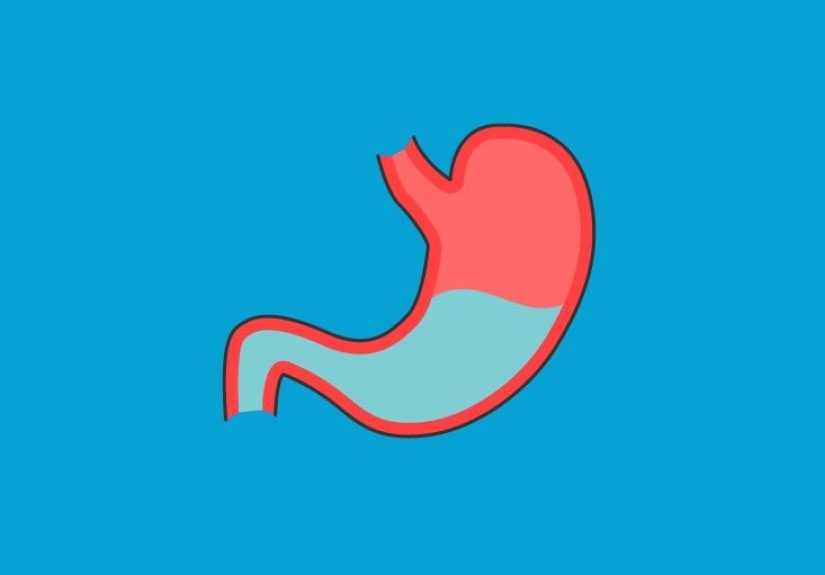
muscles to brain cells. If your stomach is the blender, the small intestine is the chemistry lab, the shipping
department, and the quality-control team… all in one squishy tube.
What the Small Intestine Does (In Plain English)
The small intestine’s main job is to finish digestion and absorb nutrients.
Food leaves the stomach as a partially digested slurry called chyme. The small intestine then:
- Breaks food down further using enzymes and bile.
- Absorbs nutrients and water through its inner lining.
- Moves leftovers into the large intestine to become waste.
Where It Fits in the Digestive Tract
Anatomically, the small intestine sits between the stomach and the large intestine (colon). The “doors” that
control traffic include the pyloric sphincter (stomach to duodenum) and the
ileocecal valve (ileum to large intestine). These valves help regulate flow and help limit
backwashbecause no one wants their intestines operating like a two-way street during rush hour.
Small Intestine Anatomy: The 3 Main Parts
The small intestine has three sectionsduodenum, jejunum, and
ileum. Each has a slightly different “specialty,” but they work as a team.
| Section | What It’s Known For | Key Highlights |
|---|---|---|
| Duodenum | Mixing & chemical digestion kickoff | Receives bile and pancreatic enzymes; helps neutralize stomach acid |
| Jejunum | Heavy-duty absorption | Absorbs many carbs, proteins, fats, vitamins, and minerals |
| Ileum | Final absorption + immune support | Absorbs vitamin B12 and bile salts; contains many Peyer’s patches |
1) Duodenum: The “Mixing Bowl” That Means Business
The duodenum is the first segment right after the stomach. It’s short compared with the rest
of the small intestine, but it’s a big deal because it’s where your body pours in the important “helpers”:
bile (from the liver/gallbladder) and digestive enzymes (from the pancreas).
Bile helps break fats into smaller droplets (emulsification), and enzymes start chopping up fats, proteins,
and carbohydrates into absorbable pieces.
The duodenum also helps protect downstream intestines by reducing the acidity of stomach contents. In other words:
it takes the stomach’s “acidic chaos” and starts turning it into a more manageable chemistry experiment.
2) Jejunum: The Absorption MVP
The jejunum is the middle portion and the workhorse for absorption. Its lining is built for
maximum contact with food, which helps your body absorb a large share of nutrients efficiently.
If digestion is a group project, the jejunum is the person doing 70% of the work while everyone else “checks email.”
3) Ileum: The Closer (With a Side Job in Immunity)
The ileum is the final section, connecting to the large intestine at the ileocecal valve.
It continues absorption and is especially important for absorbing vitamin B12 and
bile salts (which your body recycles to reuse for fat digestion later). The ileum also contains
many Peyer’s patchesclusters of immune tissue that help monitor gut contents and support immune defense.
The “Wall” of the Small Intestine: Layers That Do Different Jobs
The small intestine isn’t just an empty tubeit’s built like a multi-layered piece of high-tech clothing:
each layer has a specific job.
Key Layers (Outside to Inside)
- Serosa: A smooth outer covering that helps reduce friction with surrounding organs.
- Muscularis: Muscle layers that churn and move food along (motility).
- Submucosa: Support layer with blood vessels, lymph vessels, and nerves.
- Mucosa: The inner lining where absorption and secretion happen.
Important Cells You’ll Hear About
- Enterocytes: Absorptive cellsthe main nutrient “gatekeepers.”
- Goblet cells: Produce mucus to protect and lubricate.
- Paneth cells: Help defend against harmful microbes.
- Enteroendocrine cells: Release hormones that help regulate digestion and movement.
How the Small Intestine Moves Food: Segmentation & Peristalsis
Two main movement patterns keep digestion efficient:
- Segmentation: Back-and-forth mixing motions that churn chyme so enzymes can work and the lining can absorb nutrients.
- Peristalsis: Wave-like contractions that push contents forward through the intestine.
Much of this motion is coordinated by the enteric nervous system, sometimes called the “second brain”
of the gut because it can manage many digestive actions without you consciously thinking about it.
Absorption Superpowers: Folds, Villi, and Microvilli
Absorption is all about surface area. The small intestine uses a “triple upgrade” to maximize contact
with nutrients:
1) Circular folds (plicae)
Large ridges in the lining that slow down chyme and increase surface area.
2) Villi
Finger-like projections on the mucosal surface that absorb nutrients. Each villus contains tiny blood vessels
(capillaries) and a lymph vessel (a lacteal) for transporting absorbed nutrients.
3) Microvilli (Brush Border)
Even smaller projections on the surface of enterocytes. Microvilli massively expand surface area and hold
brush border enzymes that finish digesting carbohydrates and proteins right at the absorption site.
What Gets Absorbed Where (Examples You Can Actually Picture)
While there’s overlap, different nutrients tend to have “favorite routes” into your body:
Carbohydrates
Complex carbs are broken down into simple sugars (like glucose). These are absorbed into blood vessels and carried
to the liver through the portal circulation.
Proteins
Proteins are digested into amino acids and small peptides, which are absorbed into blood and used for building and repair.
Fats
Fats are emulsified by bile and digested into fatty acids and monoglycerides. Many fat products enter the
lacteals (lymphatic vessels) before eventually reaching the bloodstreambasically taking the “scenic route.”
Vitamin B12 and bile salts
These are especially tied to the ileum. Vitamin B12 absorption is complex and depends on other
digestive steps (like binding to intrinsic factor made in the stomach), but the ileum is the key absorption site.
Water and electrolytes
The small intestine also absorbs a large amount of water and electrolytes, supporting fluid balance.
Blood Supply, Lymphatics, and the “Shipping System”
Once nutrients cross the intestinal lining, they must be transported efficiently:
- Most sugars and amino acids enter blood capillaries and travel to the liver via portal circulation.
- Many fats enter lymph vessels (lacteals) first, then later drain into the bloodstream.
This split system helps your body manage different nutrient types and process them in the right order.
Small Intestine Diagram (Simple, Copy-Friendly)
Gross Anatomy Diagram
Microanatomy “Zoom-In” Diagram
Why Small Intestine Function Matters: Common Problems (High-Level Overview)
When the small intestine is inflamed, damaged, blocked, or missing (after surgery), the body may struggle to absorb nutrients.
That can lead to symptoms such as persistent diarrhea, abdominal pain, bloating, fatigue, and unintended weight loss.
Examples of conditions that can affect the small intestine
- Celiac disease: immune reaction can damage villi and reduce absorption.
- Crohn’s disease: inflammatory bowel disease that can affect the small intestine (often the ileum).
- Small intestinal bacterial overgrowth (SIBO): excess bacteria can interfere with digestion and absorption.
- Obstruction: scar tissue, hernias, or other issues can block passage of contents.
- Short bowel syndrome: reduced intestine length can cause malabsorption after surgery.
If someone has ongoing symptomsespecially blood in stool, severe pain, dehydration signs, or unexplained weight lossthose are reasons
to seek medical evaluation rather than trying to “DIY” the digestive system.
How to Support a Healthy Small Intestine (Practical, Non-Hype Basics)
- Eat a balanced diet with adequate protein, healthy fats, and micronutrient-rich foods.
- Stay hydrated, especially during illness or persistent diarrhea.
- Don’t ignore persistent symptomsearly evaluation can prevent nutrient deficiencies.
- Be cautious with frequent NSAID use (some people are more sensitive to gut irritation).
- Manage stress and sleep: the gut-brain connection is real, and symptoms can worsen under chronic stress.
Real-World Experiences Related to Small Intestine Function, Anatomy & Diagram (About )
Ask people what their small intestine feels like, and most will say: “I didn’t even know I had one… until it complained.”
That’s kind of the small intestine’s vibequiet competence when everything is fine, dramatic feedback when something is off.
One of the most common everyday experiences tied to small intestine function is the post-meal “gurgle orchestra.” That
bubbling, shifting sensation (often called borborygmi) is usually a normal sign of the intestine mixing and moving
contents along. It’s segmentation and peristalsis doing their jobsbasically the gut’s version of stirring soup and
sending it down the line.
In clinics, a frequent “aha” moment happens when patients learn how absorption actually works. Many people picture digestion
as food being “used up,” but the key is that nutrients must cross the intestinal lining. When clinicians explain villi and
microvilli, patients often immediately understand why damage matters: if you flatten the surface area, you reduce the body’s
ability to pick up nutrientslike trying to dry off with a towel that’s been folded into a tiny square. This explanation
comes up a lot in conversations about celiac disease, where people often describe a frustrating cycle of eating enough but
still feeling tired, weak, or foggy until the underlying issue is identified and treated.
Another real-world experience is the diagnostic journey. People with persistent symptoms may undergo tests like bloodwork,
breath tests (for certain malabsorption patterns), imaging, or endoscopic procedures. When the small intestine is hard to reach,
clinicians sometimes use capsule endoscopyswallowing a small camera that takes pictures as it travels. Patients often describe
this as surprisingly easy (it’s basically “take a vitamin-sized device and go about your day”), but the bigger experience is
emotional: relief at finally getting answers, and anxiety while waiting for results. Digestive symptoms can be embarrassing, and
simply being taken seriously can feel like a major turning point.
The ileum’s “special assignments” show up in lived experiences too. For example, some people who have had surgery involving the
terminal ileum later learn about vitamin B12 the hard waythrough fatigue, numbness/tingling, or lab results showing deficiency.
That often leads to a very practical routine: scheduled B12 supplementation under medical guidance. It’s a tangible reminder that
anatomy isn’t just a diagramit’s a set of responsibilities, and when a section is removed or inflamed, the workload doesn’t magically
disappear.
Even in classrooms, anatomy becomes “real” through experience. Medical and nursing students often remember jejunum vs ileum not by
memorizing a list, but by attaching a story: “The jejunum looked thicker and more folded,” or “The ileum had more immune patches.”
A diagram stops being just labels when you connect it to functionbile meeting chyme in the duodenum, nutrients crossing villi,
fats entering lacteals, and the ileocecal valve acting like a bouncer at the colon’s door. The most lasting experience is the
realization that digestion is not one eventit’s a coordinated chain of steps, and the small intestine is the busiest intersection
in the whole route.